The right pulmonary artery (RPA) is one of the branches of the pulmonary trunk, branching at the level of the transthoracic plane of Ludwig It is longer than the left pulmonary artery and courses perpendicularly away from the pulmonary trunk and left pulmonary artery, between the superior vena cava and the right main bronchusAs it courses to the right it has an almost horizontal pathBrowse 348 pulmonary artery stock photos and images available, or search for pulmonary hypertension or pulmonary embolism to find more great stock photos and pictures human heart pulmonary artery stock illustrations human anatomy, section, chambers of heart, victorian anatomical drawing pulmonary artery stock illustrations Pulmonary Veins The pulmonary veins receive oxygenated blood from the lungs, delivering it to the left side of the heart to be pumped back around the body There are four pulmonary veins, with one superior and one inferior for each of the lungs They enter the pericardium to drain into the superior left atrium, on the posterior surface

How The Heart Works The Community Cardiology Service
Pulmonary artery and vein anatomy
Pulmonary artery and vein anatomy-This video covers the anatomy, location and function of the pulmonary veins, directing the blood from lung to heart Test yourself with our quiz on the surroThe oxygenated blood is taken through the pulmonary vein back into the heart through the left atrium and the left ventricle It is then pumped throughout the body Anatomy The pulmonary artery is an extension of the pulmonary trunk (lat truncus pulmonaris), extending from the right ventricle;
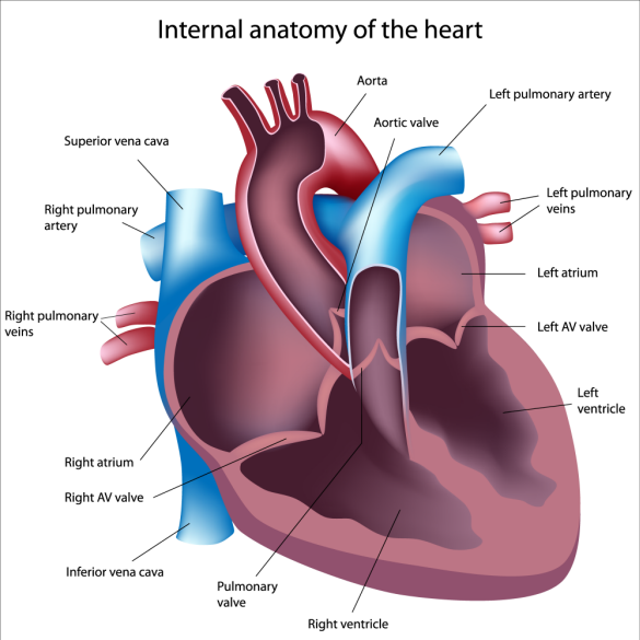


Echocardiography And Pulmonary Arterial Hypertension A Detailed Look
LEFT PULMONARY ARTERYTheleft pulmonary artery, arising in front of the left main bronchus, ascends slightly in a posterior and lateral direction, arches over the left main bronchus, and then hooks round the left upper lobe bronchus, following a subpleural course throughout (Figs V, VI, and VII) It more fully establishes its superiority over the left superior pulmonary vein (Fig VIII) Pulmonary Vein The segmental and subsegmental arteries of the pulmonary vein run independently from the bronchi in the interlobular septa Anatomy of the Wall Pulmonary Artery Pulmonary artery wall is thick and elastic Pulmonary Vein Pulmonary vein wall is thin when compared to the pulmonary artery ValvesCT scan shows an enlarged main pulmonary artery (MPA) that measures 51cms at the level of the tubular portion of the ascending aorta 71 year old female with long standing idiopathic pulmonary hypertension, and retroperitoneal fibrosis The MPA is enlarged The pulmonary valve is normal in thickness, thus excluding pulmonary stenosis
Ecr 07 C 164 Three Dimensional Ct Angiography Of Various Hello, What's up guys?Pulmonary Arteries and Veins Variant Image ID 6476 Add to Lightbox Save to Lightbox Email this page Link this page Print Please describe! Pathology of the pulmonary vasculature involves an impressive array of both congenital and acquired conditions While some of these disorders are benign, disruption of the pulmonary vasculature is often incompatible with life, making these conditions critical to identify on imaging Many reviews of pulmonary vascular pathology approach the pulmonary arteries, pulmonary veins
I am so proud to present you today thisOur LATEST youtube film is ready to run Just need a glimpse, leave your valuable advice let us know , and subscribe us! The pulmonary vein in the interlobular septa join together to form segmental pulmonary veins Unlike the segmental pulmonary arteries, the segmental pulmonary veins are not close to the bronchi, instead they run within the intersegmental septa The segmental pulmonary veins join to form two common trunks of the superior pulmonary vein and the inferior pulmonary vein which connect to the left atrium on each side The orifices of the inferior pulmonary veins


Echocardiography And Pulmonary Arterial Hypertension A Detailed Look


References In Pulmonary Vascular System And Pulmonary Hilum Thoracic Surgery Clinics
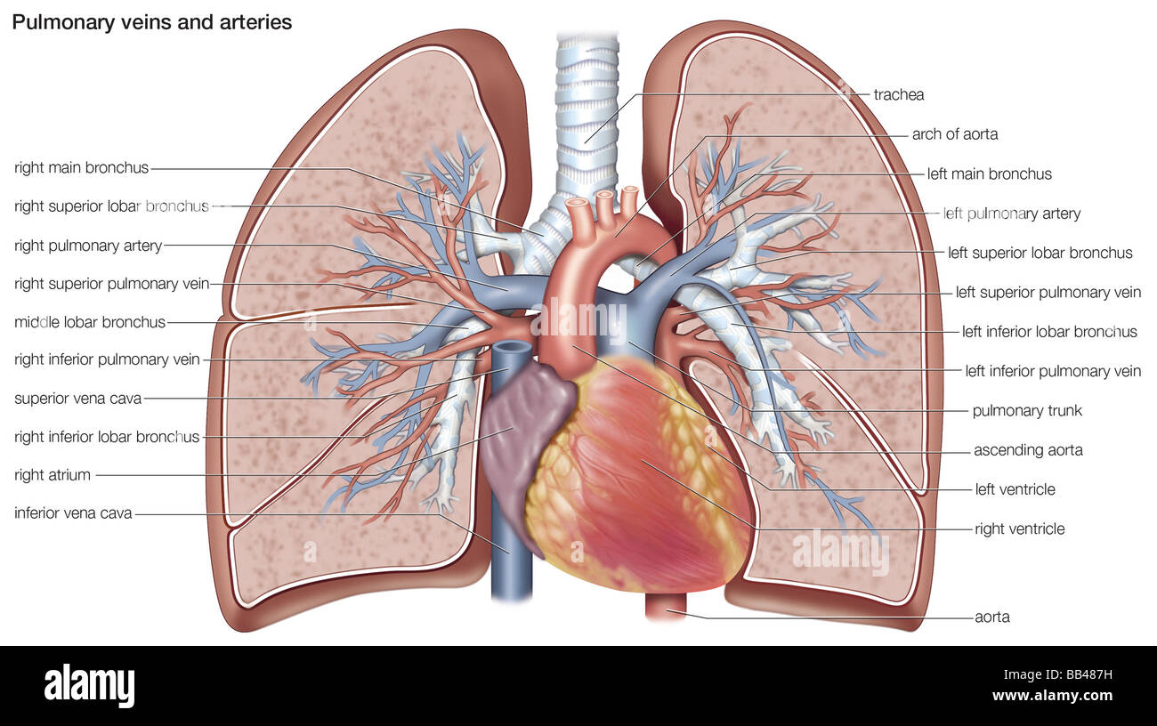
Pulmonary Arteries The main pulmonary artery arises from the right ventricle distal to the pulmonary valve and courses cephalad and dorsally; The pulmonary arteries and the pulmonary veins are the vessels of the pulmonary circulation;A pulmonary artery is an artery in the pulmonary circulation that carries deoxygenated blood from the right side of the heart to the lungs The largest pulmonary artery is the main pulmonary artery or pulmonary trunk from the heart, and the smallest ones are the arterioles, which lead to the capillaries that surround the pulmonary alveoli


Skill Lab Learning


Pulmonary Artery Anatomy Britannica
Which means they are responsible for carrying the oxygenated blood to the heart from the lungs and carrying the deoxygenated blood from the heart to the lungsHigh quality Pulmonary Arteryinspired gifts and merchandise Tshirts, posters, stickers, home decIt divides into right and left pulmonary arteries (Figs 11, 12, 13, 14, and 15) The right pulmonary artery divides into the truncus anterior (superior trunk) and the interlobar pulmonary artery


Pulmonary Trunk Radiology Reference Article Radiopaedia Org
/heart-and-circulatory-system-with-blood-vessels--97537745-a3bc2b2a6ca94390bfdf2696ad9bbddd.jpg)


Pulmonary Vein Anatomy Function And Significance
Arteries are vessels that carry blood away from the heartThe main pulmonary artery or pulmonary trunk transports blood from the heart to the lungsWhile most major arteries branch off from the aorta, the main pulmonary artery extends from the right ventricle of the heart and branches into left and right pulmonary arteries The left and right pulmonary arteries extend toA The pulmonary arteries bring blood with less oxygen content to the lungs where the blood is enriched with oxygen and it is transported back into the heart with the help of the pulmonary veins This oxygenated blood travels into the heart's left atrium and is then pumped to the left ventricle Then it is circulated through the aorta to the arteries which carry it throughout all The anatomy of pulmonary vessels varies The right upper pulmonary vein usually drains in front of the pulmonary artery to the left atrium We herein describe a case of the right upper lobe pulmonary vein draining posterior to the pulmonary artery and absent right upper lobe pulmonary vein in the ventral hilum A 64yearold woman suspected to have lung cancer and
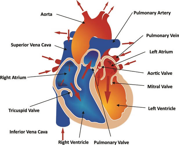


Pulmonary Artery The Definitive Guide Biology Dictionary


Airways And Lungs Knowledge Amboss
Pulmonary embolism (PE) occurs when a thrombus dislodges from a vein, flows through the veins and typically lodges in the lung Most thrombi form in one of the deep veins of the lower limb or those of the pelvis;Pulmonary veins The smaller pulmonary veins are mainly distinct from the smaller pulmonary arteries by being fewer in number of branches (only 115 orders) and having thinner walls, which are less muscular, less elastic, and more collagenous The largest pulmonary veins flow seamlessly into the left atrium Unlike the large arteries, theseThis condition is referred to as deep vein thrombosis (DVT) The thrombi lodge in the lungs because veins get larger as they flow to


Difference Between Pulmonary Artery And Pulmonary Vein Definition Characteristics Function


Pulmonary Veins Function Definition Anatomy Video Lesson Transcript Study Com
Pulmonary arteriovenous malformations (AVMs) are pathological connections between the pulmonary artery and the pulmonary vein without an intermediary capillary network resulting in righttoleft shunt Pulmonary AVMs are typically congenital, thought to be present at birth, but typically only become clinically relevant in adulthood Hereditary haemorrhagic telangiectasiaIt is located in front and to the left of all the vessels that flow in and out of the heart, On the left side, the superior left pulmonary artery drains the left upper lobe and the inferior left pulmonary artery the lower lobe In some people, the three right pulmonary veins remain separate instead of merging into two veins, resulting in a total of five pulmonary veins (this is referred to as a single accessory right middle pulmonary vein and is present in roughly 10% of
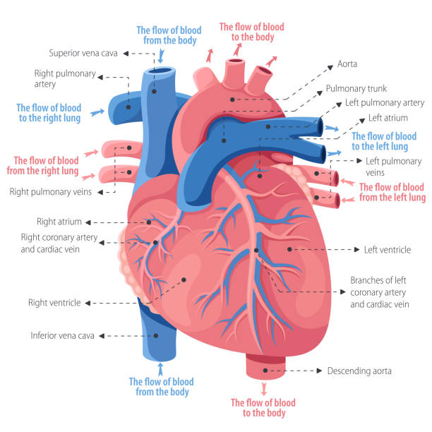


671 Pulmonary Vein Stock Photos Pictures Royalty Free Images Istock
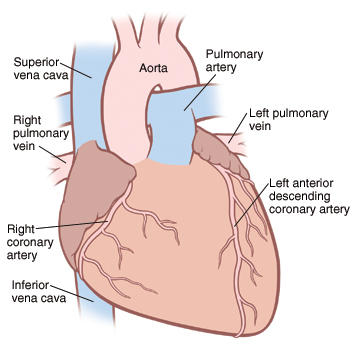


How The Heart Works Saint Luke S Health System
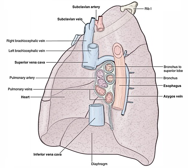
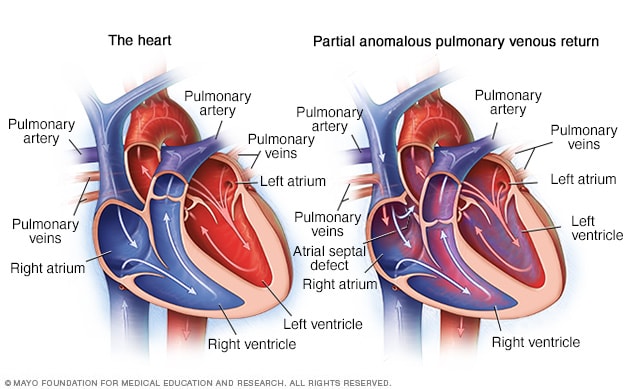
The Anatomy of the Pulmonary Vein The deoxygenated blood that is carried back from the periphery of the body in venules and veins reaches the Normal anatomy of pulmonary veins The blue (deoxygenated) marks pulmonary arteries, and the red (oxygenated) marks the pulmonary veins In atypical but not rare situations in human, pulmonary veins that both originate from right (4%) or left (178%) may fuse into a common trunk before entering the left atrium Additional pulmonary veins derive from individualObjectives The size of the pulmonary veins (PVs) and pulmonary arteries (PAs) changes in response to hemodynamic alterations caused by physiological events and disease We sought to create standardized echocardiographic methods for imaging the right ostium of the pulmonary veins (RPVs) and the right pulmonary artery (RPA) using specific landmarks and timing to


Pulmonary Veins Radiology Reference Article Radiopaedia Org
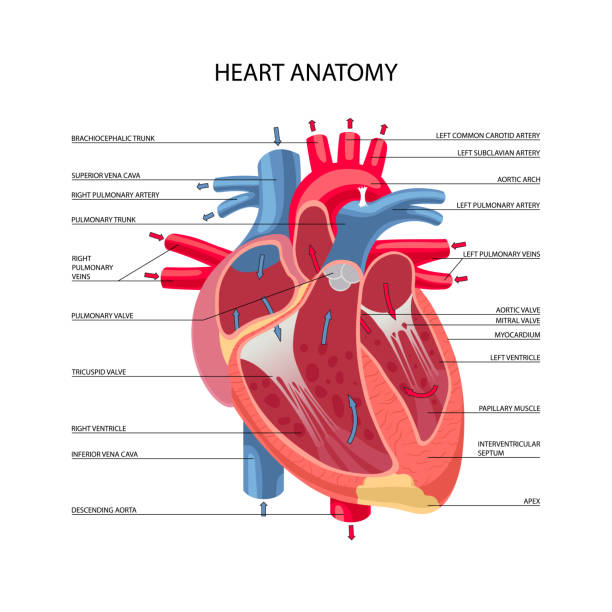


1 590 Pulmonary Artery Stock Photos Pictures Royalty Free Images Istock
We are pleased to provide you with the picture named Pulmonary Lung Artery And Vein AnatomyWe hope this picture Pulmonary Lung Artery And Vein Anatomy can help you study and research for more anatomy content please follow us and The vessels supplying the lungs include the pulmonary arteries, pulmonary veins, and bronchial arteries The segmental and sub segmental pulmonary arteries parallel the bronchi and are named according to the bronchopulmonary segments they supply There are however considerable anatomic variations, particularly in the upper lobes with variations in number or presence of accessory arteries from adjacent segments The subsegmental pulmonary vein The pulmonary vein (D) divides again, developing its typical 2 left and 2 right pulmonary veins as well as the progressive network of major venous channels paralleling the arterial, lymphatic, and bronchial channels Within the lung parenchyma proper, the pulmonary vasculature remains underdeveloped until the end of the canalicular period (∼26 weeks), before


The Pulmonary Trunk


1
How you will use this image and then you will be able to add this image to your shopping basket Portal System, Pulmonary & Systemic Veins Leave a Comment / Staff Nurse / Anatomy and physiology MCQs for nurses to prepare all type of government exams, nursing school andANGEL biology classes bhatpar rani


The Pulmonary Artery Carries Blood To Lung For Blood Oxidation And Renal Artery Carries Blood To Kidney For Blood Purification How Can The Lungs And Kidneys Get Oxygen And Food Is It


Pictorial Review Of The Pulmonary Vasculature From Arteries To Veins Insights Into Imaging Full Text
We are pleased to provide you with the picture named Pulmonary artery and vein anatomy We hope this picture Pulmonary artery and vein anatomy can help you study and research for more anatomy content please follow us and visit our website wwwanatomynotecom Anatomynotecom found Pulmonary artery and vein anatomy from plenty of anatomical The vessels supplying the lungs include the pulmonary arteries, pulmonary veins, and bronchial arteries The segmental and sub segmental pulmonary arteries parallel the bronchi and are named according to the bronchopulmonary segments they supply There are however considerable anatomic variations, particularly in the upper lobes with variations in number or presence of accessory arteries from adjacent segments The subsegmental pulmonary veinWe've gathered our favorite ideas for Pulmonary Artery Anatomy Ct, Explore our list of popular images of Pulmonary Artery Anatomy Ct and Download Photos Collection with high resolution


Pulmonary Hypertension Symptoms Classes Medications Life Expectancy


348 Pulmonary Artery Photos And Premium High Res Pictures Getty Images
Footnote This figure includes six CT or MR images of the left atrium and pulmonary veins viewed from the posterior perspective Common and uncommon variations in pulmonary vein anatomy are shown (A) Standard pulmonary vein anatomy with 4 distinct pulmonary vein ostia (B) Variant pulmonary vein anatomy with a right common and a left common pulmonary veinNormal pulmonary vein anatomy is also depicted, with a hilar angle formed by the intersection of the vertically oriented superior pulmonary vein superiorly and the interlobar artery inferiorly, at the hilum On a lateral view, the left superior pulmonary vein can be seen anterior to the trachea, superimposed on the right pulmonary artery and nearly contiguous with the left pulmonary artery before it arches In cases of pulmonary venous hypertension, the superior pulmonary veinsAnatomical terminology The pulmonary veins are the veins that transfer oxygenated blood from the lungs to the heart The largest pulmonary veins are the four main pulmonary veins, two from each lung that drain into the left atrium of the heart The pulmonary veins
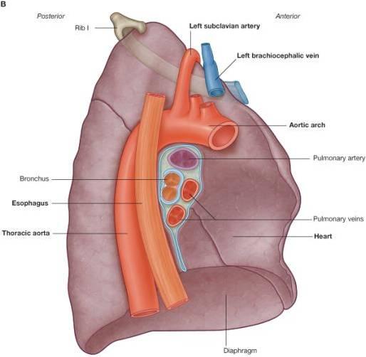


Thorax Venous Structure Pulmonary Veins Ranzcrpart1 Wiki Fandom
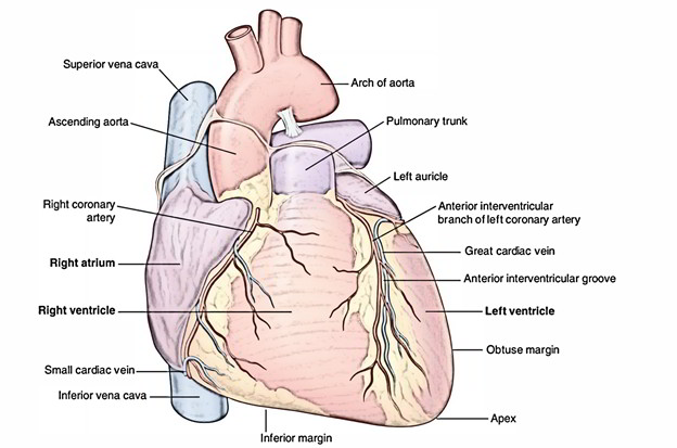


Easy Notes On Pulmonary Arteries Learn In Just 3 Minutes Earth S Lab
Gross anatomy There are typically four pulmonary veins, two draining each lung right superior drains the right upper and middle lobes right inferior drains the right lower lobe left superior drains the left upper lobe left inferior drains the left lower lobe The single pulmonary trunk that lies behind the pulmonary valve splits into right and left pulmonary arteries that are elastic when healthy When this elasticity is lost, pulmonary arterial hypertension can occur Pulmonary Artery Anatomy The main pulmonary artery exits the heart above the pulmonary valve of the right ventricle This pulmonary trunk has a length ofOther articles where Pulmonary vein is discussed pulmonary circulation and larger vessels until the pulmonary veins (usually four in number, each serving a whole lobe of the lung) are reached The pulmonary veins open into the left atrium of the heart Compare systemic circulation


Pulmonary Arteries Location Function Human Anatomy Kenhub Youtube


Siology 15th Edition 125 Right Pulmonary Artery Left Chegg Com
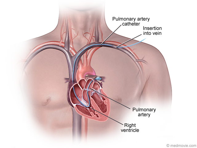
Pulmonary Artery Anatomy The main pulmonary artery, also known as the pulmonary trunk, originates at the right ventricle at the point of the pulmonary valve, a oneway semilunar valve that allowsRight superior pulmonary vein Pulmonary artery catheter Middle lobe pulmonary vein Left atrium Right inferior pulmonary vein First of 2 images from a normal pulmonary arteriogram shows the normal anatomy of the pulmonary veins AP view of the venous phase of a right pulmonary arteriogram shows an arterial catheter that courses through the right atrium, right ventricle, pulmonary Pulmonary vein isolation (PVI) over the last decade has become the most demanded method for AF treatment Even with experience in ablation, knowledge of left atrial anatomy and pulmonary vein anatomy is necessary A detailed visualization of the left atrium (LA) and pulmonary vein (PV) anatomy can be obtained by several different imaging


Pulmonary Arteries Location Function Human Anatomy Kenhub Youtube


Pulmonary Arteries And Veins


Pulmonary Arteries Veins Arteries And Veins Carotid Artery Arteries


Bronchus Pulmonary Artery Veins Model Welcome


Pulmonary Artery And Vein Anatomy


Pulmonary Arteries Stock Illustrations 954 Pulmonary Arteries Stock Illustrations Vectors Clipart Dreamstime
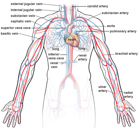


Illustrations Of The Blood Vessels


Livingwithph Ca Blood Flow In The Pulmonary Arteries Youtube
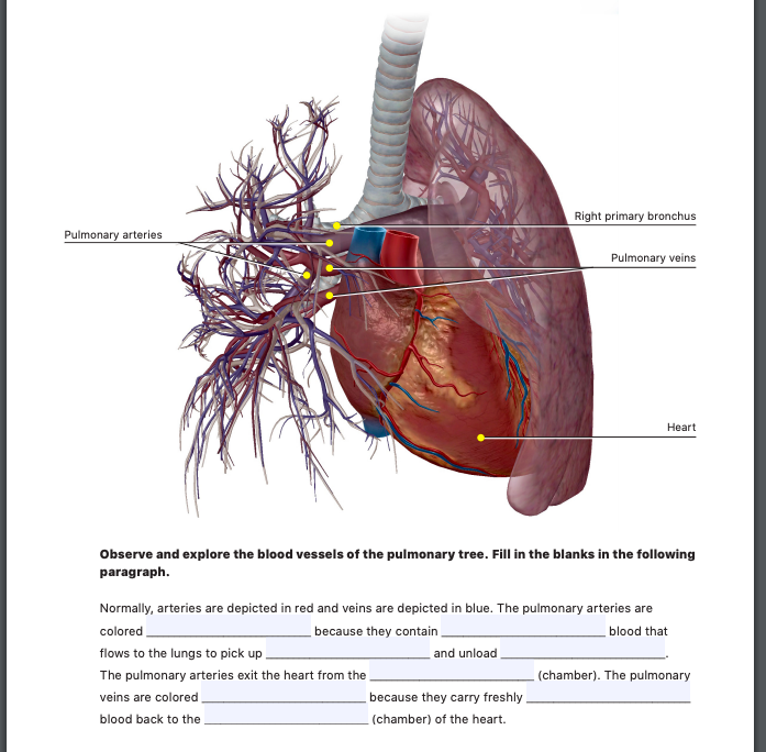


Right Primary Bronchus Pulmonary Arteries Pulmonary Chegg Com
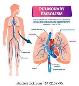


Pulmonary Veins Images Stock Photos Vectors Shutterstock


Right Pulmonary Artery Radiology Reference Article Radiopaedia Org
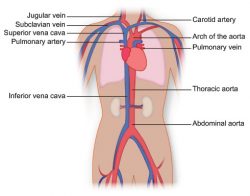


Vasculature Of The Torso Texas Heart Institute


What Is Special About The Pulmonary Artery And Pulmonary Vein Quora


Easy Notes On Pulmonary Veins Learn In Just 4 Minutes Earth S Lab


Partial Anomalous Pulmonary Venous Return Overview Mayo Clinic


Pulmonary Artery Catheter


Pulmonary Veins And Arteries Stock Photo Alamy
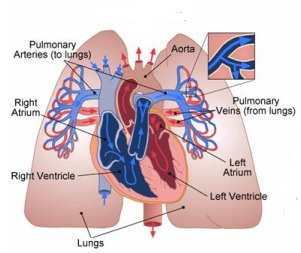


What Does The Pulmonary Artery Do Socratic
:watermark(/images/watermark_only.png,0,0,0):watermark(/images/logo_url.png,-10,-10,0):format(jpeg)/images/anatomy_term/pulmonary-artery-3/AnLPQFdAn7BHBaVWuILOw_Pulmonary_arteries_-2-_.png)


Pulmonary Arteries And Veins Anatomy And Function Kenhub
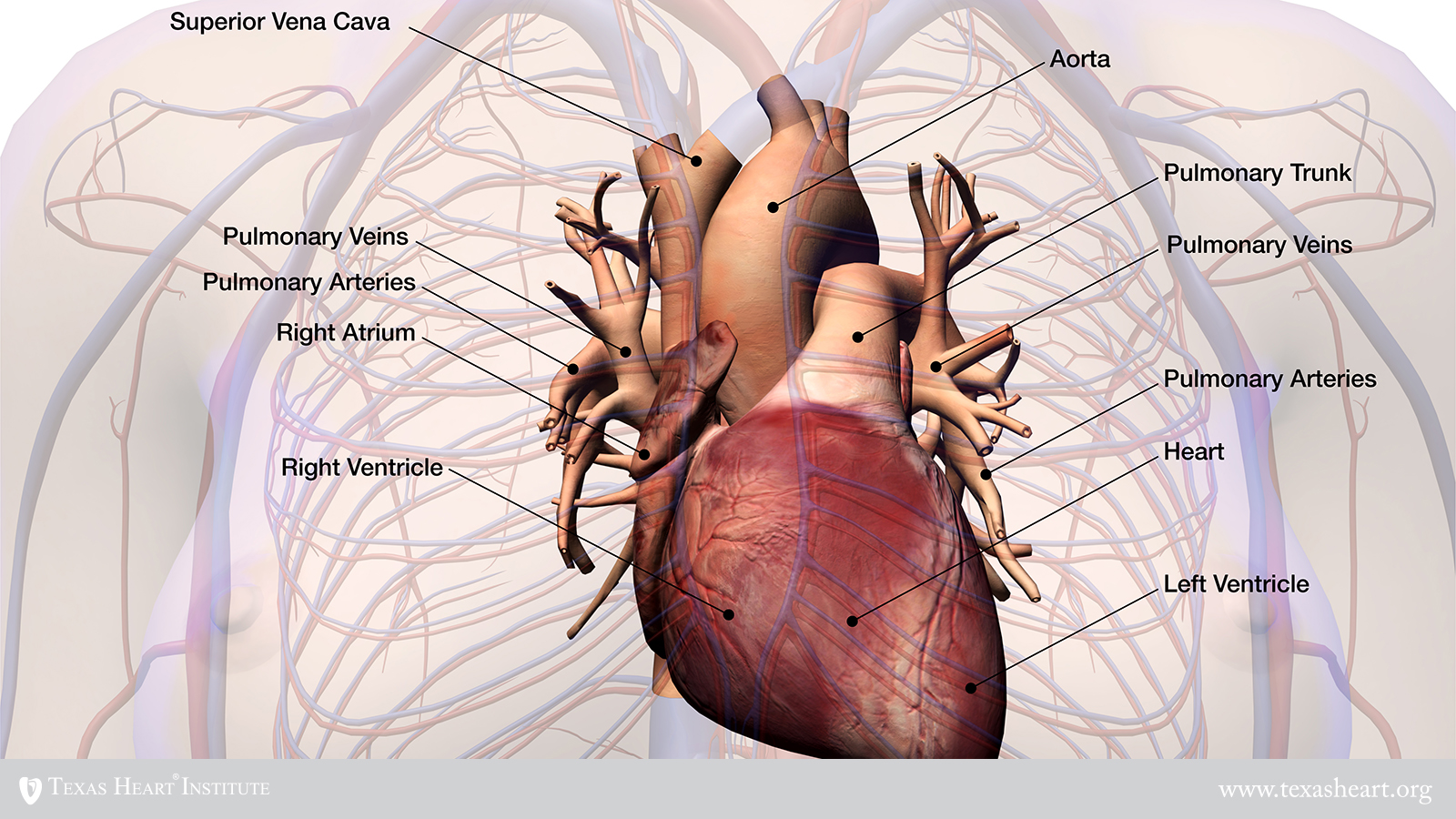


Transposition Of The Great Arteries Texas Heart Institute


Hilum Anatomy Britannica
:background_color(FFFFFF):format(jpeg)/images/library/7779/bronchioles-alveoli-anatomy_english.jpg)


Pulmonary Arteries And Veins Anatomy And Function Kenhub


152 Aorta Human Cardiovascular System Pulmonary Arteries Pulmonary Vein Stock Photos Pictures Royalty Free Images Istock


Vein Wikipedia
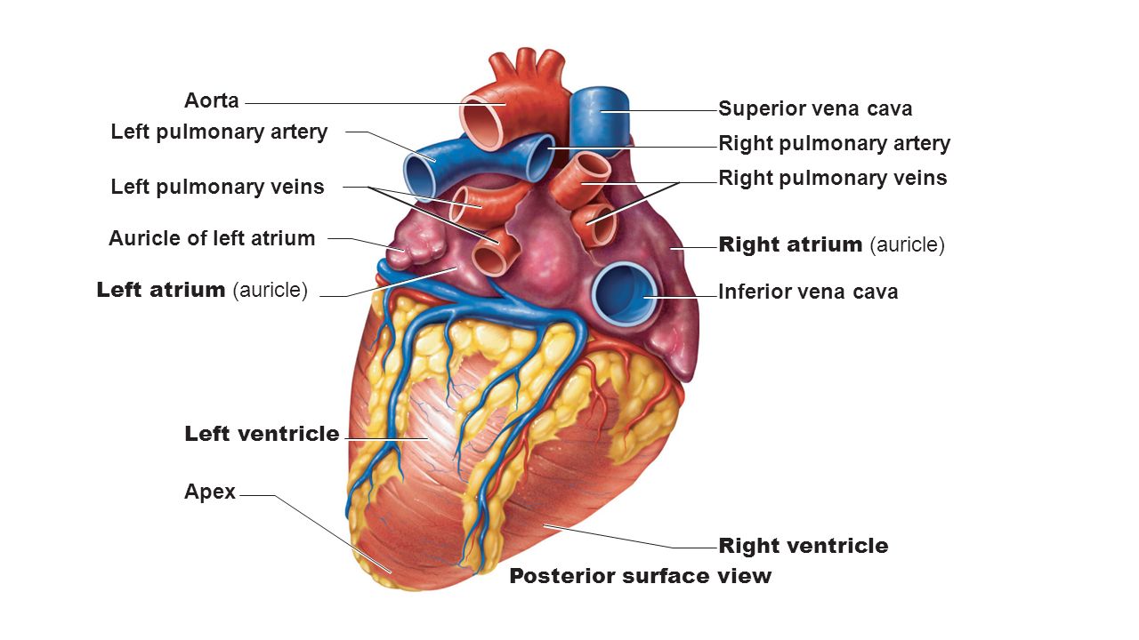


What Does The Pulmonary Vein Do Socratic
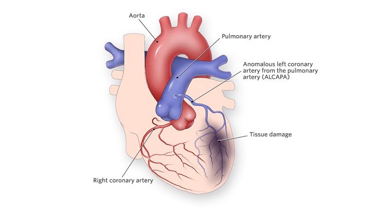


Anomalous Left Coronary Artery From The Pulmonary Artery Children S Hospital Of Philadelphia
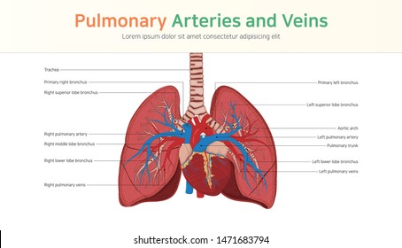


Pulmonary Veins Images Stock Photos Vectors Shutterstock


Lung Pulmonary Veins And Arteries Royalty Free Vector Image


How The Heart Works The Community Cardiology Service
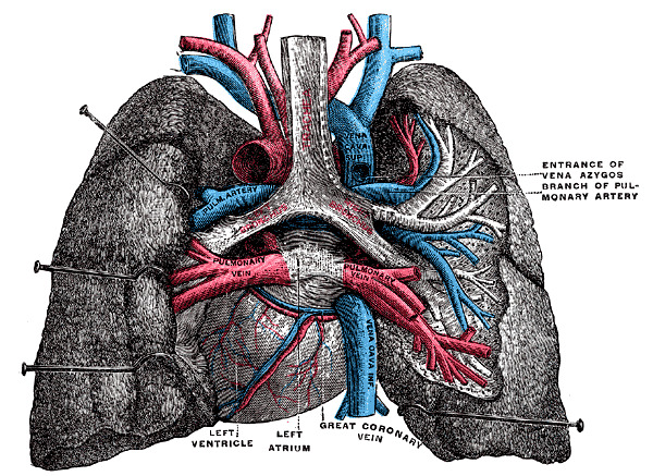


Figure Posterior View Of Heart And Statpearls Ncbi Bookshelf


Pulmonary Circulation Wikipedia


Anatomy Of The Pulmonary And Bronchial Circulation Deranged Physiology


The Circulatory System Before And After Birth
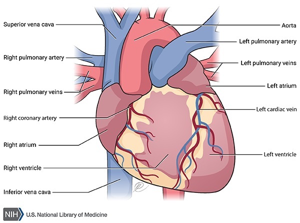


Pulmonary Veno Occlusive Disease Medlineplus Genetics
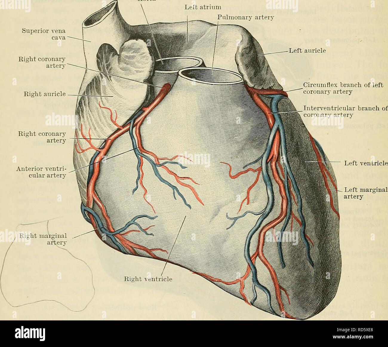


Cunningham S Text Book Of Anatomy Anatomy 872 The Vascular System The Base Is Limited Below By The Inferior Part Of The Coronary Sulcus In Which The Coronary Sinus Lies Its Upper Border


348 Pulmonary Artery Photos And Premium High Res Pictures Getty Images


Anatomy Of Pulmonary Arteries Anatomy Drawing Diagram
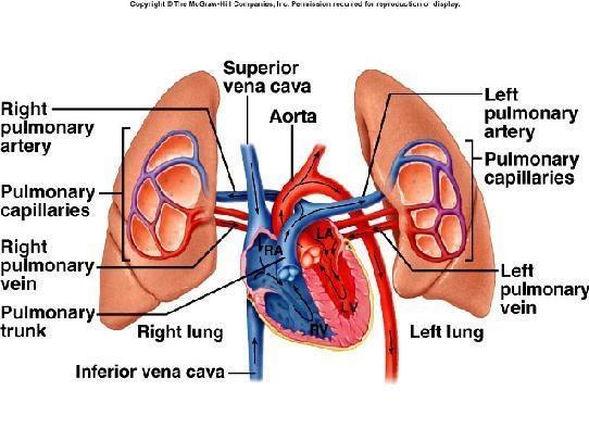


Why Pulmonary Artery Carry Deoxygenated Blood Socratic
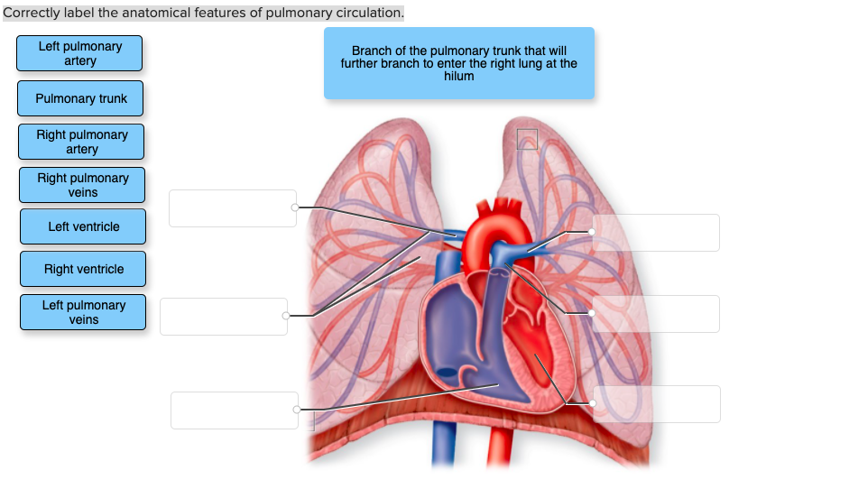


Question Correctly Label The Anatomical Features Of Pulmonary Circulation Left Pulmonary Artery Branch Of The Pulmonary Trunk That Will Further Branch To Enter The Right Lung At The Hilum Pulmonary Trunk Right
/heart-and-circulatory-system-with-blood-vessels--97537745-a3bc2b2a6ca94390bfdf2696ad9bbddd.jpg)


Pulmonary Vein Anatomy Function And Significance
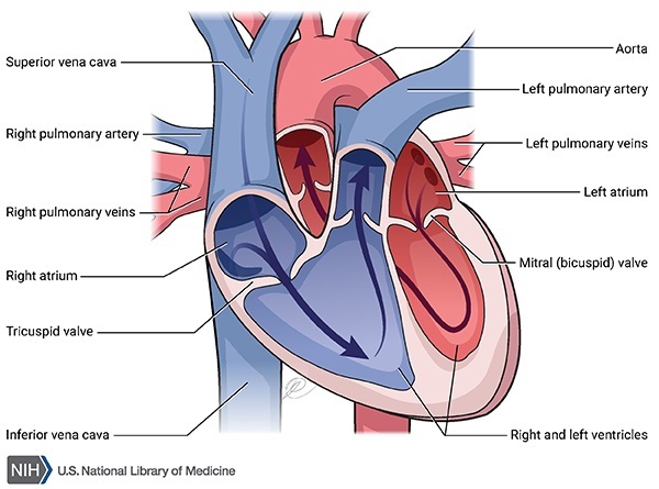


Pulmonary Arterial Hypertension Medlineplus Genetics


Pulmonary Artery Wikipedia


1


Pulmonary Vein The Definitive Guide Biology Dictionary
:max_bytes(150000):strip_icc()/GettyImages-87394349-568952ca3df78ccc152e5be5.jpg)


Pulmonary Artery Anatomy Function And Significance


Pulmonary Veins Stock Illustrations 941 Pulmonary Veins Stock Illustrations Vectors Clipart Dreamstime
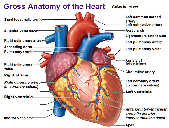


Heart Anatomy


Pulmonary Arteries Function Anatomy Video Lesson Transcript Study Com


What Is The Difference Between Pulmonary Artery And Other Arteries Pediaa Com


Pulmonary Artery Catheterization Procedures Consult
/human-heart-circulatory-system-598167278-5c48d4d2c9e77c0001a577d4.jpg)


Av And Semilunar Heart Valves


Human Being Anatomy Blood Circulation Principal Veins And Arteries Image Visual Dictionary
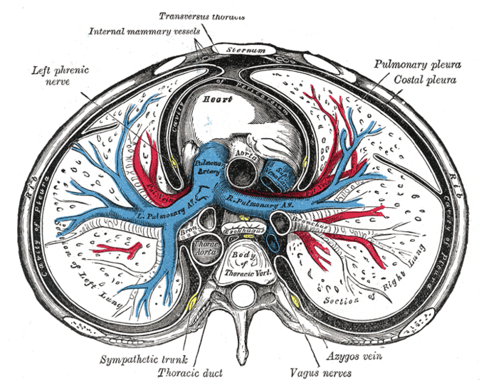


The Heart Boundless Anatomy And Physiology
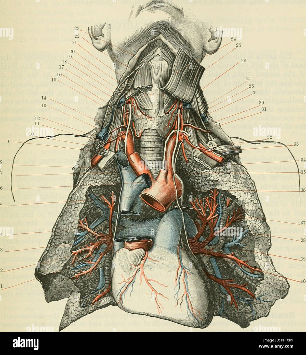


Pulmonary Arteries High Resolution Stock Photography And Images Alamy


Illustration Showing The Origin Of The Pulmonary Artery Wedge Blood Download Scientific Diagram


Exercise 45 Anatomy Of The Heart Superior Aorta Chegg Com
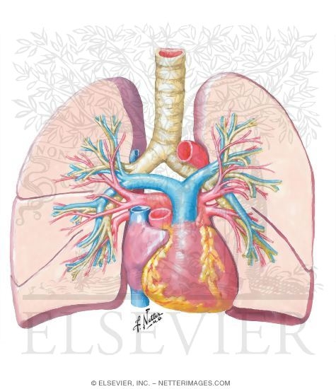


Pulmonary Arteries And Veins


Pulmonary Vein Anatomy Function Location Ablation Stenosis Thrombosis
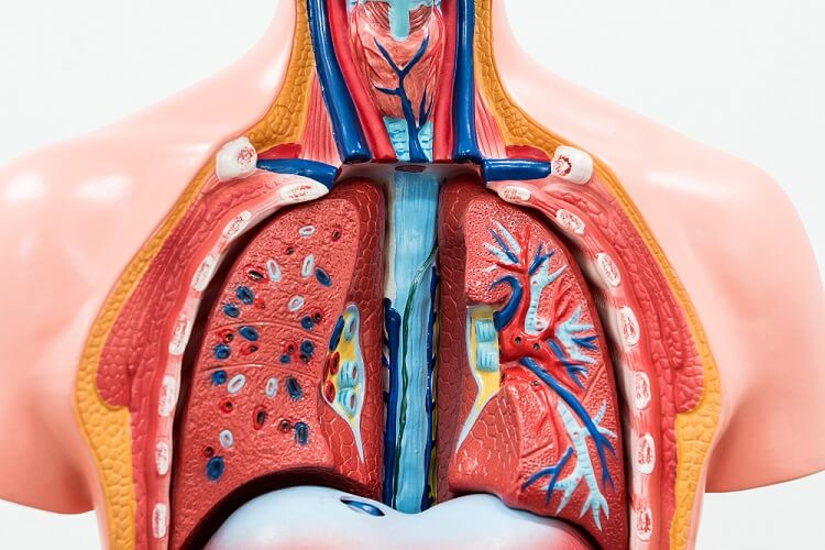


Pulmonary Artery The Definitive Guide Biology Dictionary


What Is The Pulmonary Artery Mvp Resource
/heart-lungs-5be35c5446e0fb00519cde59.jpg)


How The Main Pulmonary Artery Delivers Blood To The Lungs


Pulmonary Artery Wikipedia


Difference Between Artery And Vein
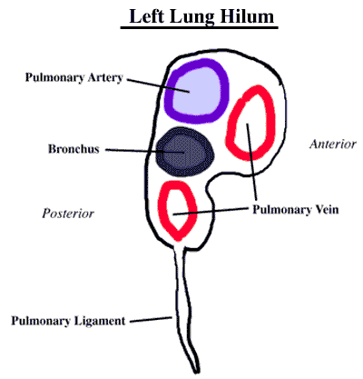


Dissector Answers Superior Mediastinum Lungs


If Blood Is In The Pulmonary Artery Does It Have More Oxygen Or More Carbon Dioxide Quora


Physiologic Anatomy Of The Pulmonary Circulatory System


Difference Between Pulmonary Artery And Pulmonary Vein Knowswhy Com


1


Main Bronchi With Pulmonary Arteries And Veins In Situ Kedokteran Medis Biologi


Pulmonary Artery Anatomy Britannica


03x Normal Pulmonary Circulation Anatomy Exhibits
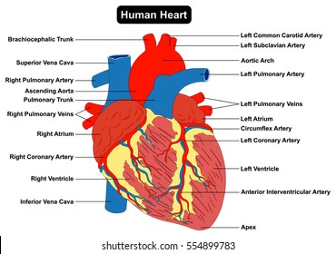


Pulmonary Veins Images Stock Photos Vectors Shutterstock


The Pulmonary Veins And Arteries In The Human Pulmonary Medical Illustration Arteries


1
:background_color(FFFFFF):format(jpeg)/images/library/7777/sternocostal-surface-of-the-heart_english.jpg)


Pulmonary Arteries And Veins Anatomy And Function Kenhub
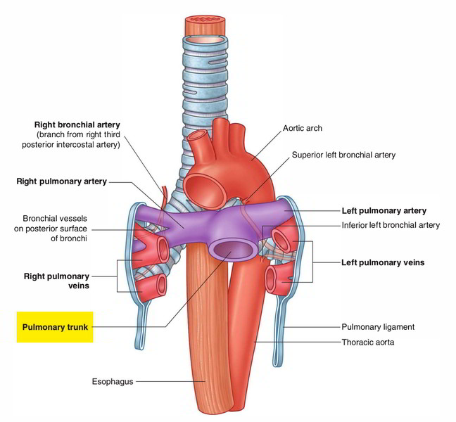


Easy Notes On Pulmonary Trunk Learn In Just 3 Minutes Earth S Lab


Congenital Heart Defects Facts About Tavpr Cdc


Congenital Defects Tutorial Congenital Heart Defects Atlas Of Human Cardiac Anatomy
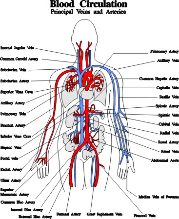


Blood Vessels Arteries Capillaries Veins Vena Cava Central Veins Lhsc
:watermark(/images/watermark_only.png,0,0,0):watermark(/images/logo_url.png,-10,-10,0):format(jpeg)/images/anatomy_term/arteria-pulmonalis-dextra-2/KXB9skOlZkZElGhw8KHrA_A._pulmonalis_dextra_01.png)


Pulmonary Arteries And Veins Anatomy And Function Kenhub


0 件のコメント:
コメントを投稿